Disclosure(s): No financial relationships with ineligible companies to disclose
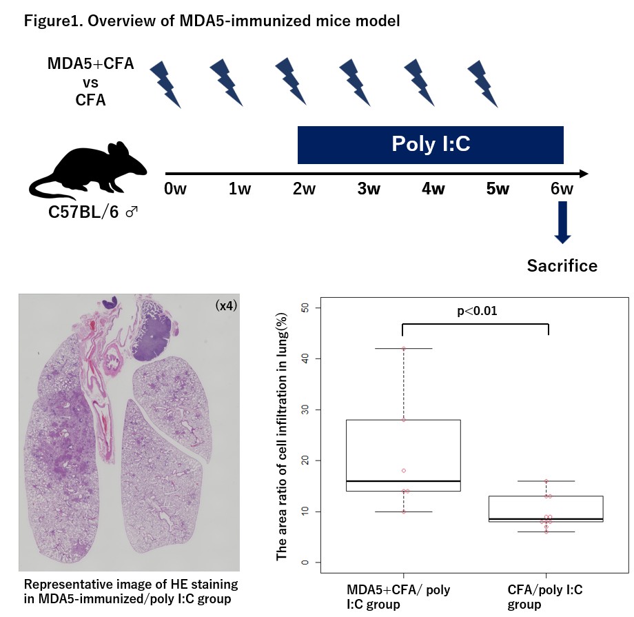
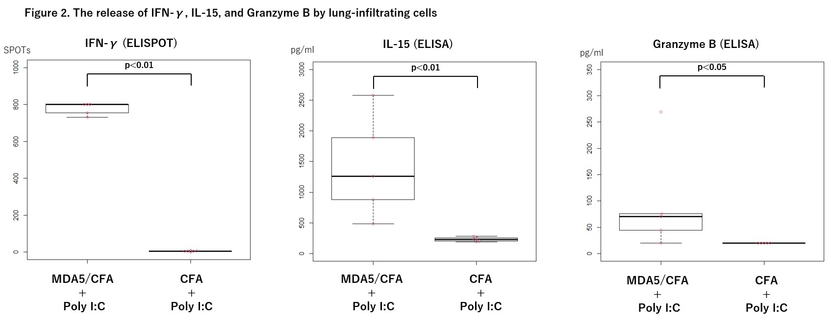
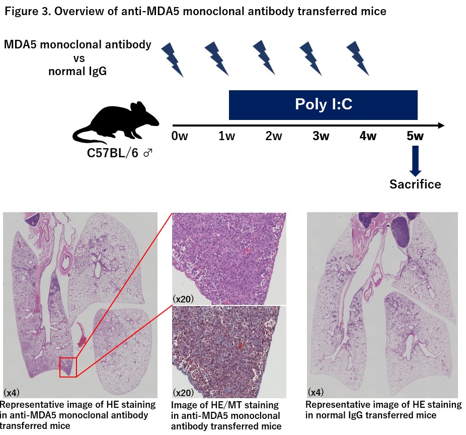
Background/Purpose: Anti-Melanoma differentiation-associated gene 5-positive dermatomyositis (MDA5-DM) is often complicated by rapidly progressive interstitial lung disease (RP-ILD) and has a poor prognosis. Although there has been one report on this disease mouse model, the role of autoantigen-specific reactivity, especially anti-MDA5 antibody itself, on the pathogenesis of the disease has never been elucidated. In this study, we established a new mouse model of MDA5-DM-ILD and investigated the meanings of autoreactivity to MDA5 in the pathogenesis of the disease. Methods: C57BL/6J wild-type mice were immunized with complete Freund’s adjuvant (CFA) and recombinant human MDA5 protein purified from baculovirus-insect cell system 6 times once a week, and polyinosinic-polycytidylic acid (poly I:C) was administered intratracheally 16 times over a 4-week period, starting from the 3rd week. The production of anti-mouse MDA5 antibodies was confirmed by immunoprecipitation. The area of cell infiltration in lung was quantitatively evaluated using an all-in-one fluorescence microscope (BZ-X810), the cell ratio was evaluated by flow cytometry, and interferon-stimulated gene (ISG) was evaluated by RT-PCR. In addition, we observed whether the lung infiltrating cells were reacted with MDA5 protein and releasing cytokines and granzyme B. After then, CD4(+) T cells and B cells were sorted and co-cultured, and IFN-γ production was confirmed by adding MDA5 protein. Furthermore, anti-human MDA5 monoclonal mouse antibodies were transferred intraperitoneally to C57BL/6J wild-type mice 5 times once a week, and poly I:C was administered intratracheally 16 times over a 4-week period, and the area of cell infiltration in lung tissue was quantitatively evaluated. Results: We confirmed the production of anti-mouse MDA5 antibodies in immunized mice. The MDA5-immunized/poly I:C mice showed larger cellular infiltration area of the lungs (p< 0.01, Figure1) and lower survival (p< 0.05) compared to CFA only immunized/poly I:C mice. ISG15 expression in lung tissue was increased. In vitro cytokine releasing assay using MDA5 protein revealed that lung-infiltrating cells of MDA5-immunized/poly I:C mice produced significantly more IFN-γ, IL-15, and granzyme B than control mice (p< 0.05, Figure2), which can be observed even after purification of infiltrating B cells and CD4(+) Tcells. The area of pneumonia in the mice injected with the monoclonal antibody was different from that in the mice injected with normal IgG, with dominant cellular infiltration just below the pleura, which was similar to the initial development of ILD in MDA5-DM patients (Figure3). Conclusion: Infiltration of MDA5-specific T/B cells in the lungs as well as anti-MDA5 antibody itself may play a pivotal role for lung injury in MDA5-DM.