University of Michigan Ann Arbor, Michigan, United States
Disclosure information not submitted.
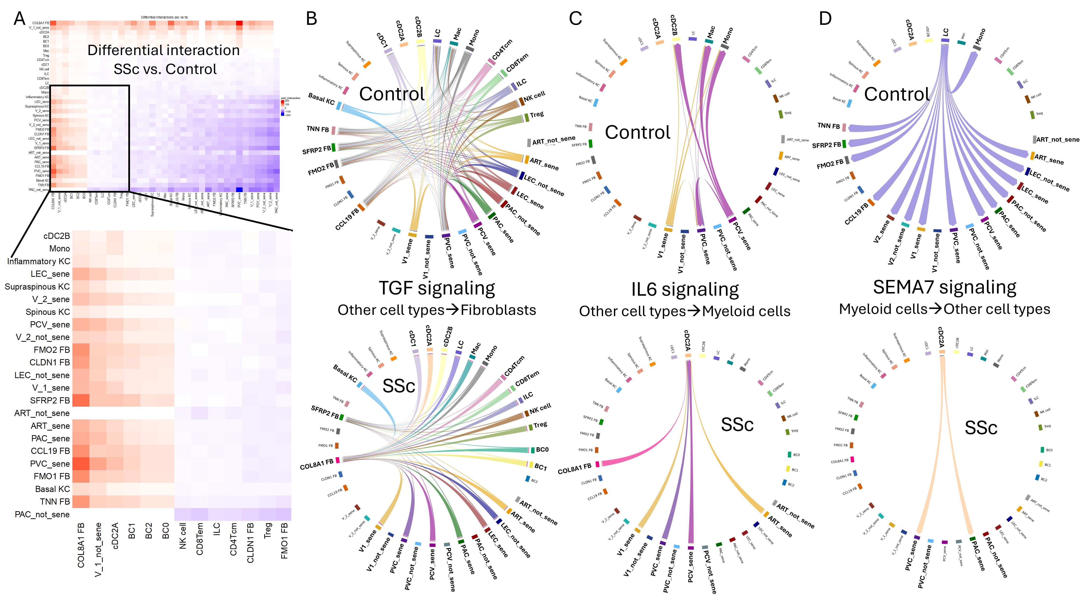
Background/Purpose: Systemic sclerosis (SSc) is characterized by extensive damage of the microvessels in multiple organs. We and others showed that endothelial cells (ECs) isolated from SSc skin biopsies show dysregulated phenotypes including impaired angiogenesis, barrier dysfunction, and spontaneous/constitutive endothelial-to-mesenchymal transition (EndoMT). Notably, these are hallmarks of EC senescence. In this study, we therefore sought to determine the presence and role of EC senescence in SSc skin fibrosis. Methods: Dermal microvascular ECs were isolated from skin biopsies of age-matched healthy individuals and SSc patients. Cellular senescence was assessed through qPCR, immunofluorescence, β-galactosidase expression and measuring the senescence-associated secretory phenotype (SASP). To investigate EC-specific senescence at the single-cell level, we analyzed skin biopsies from 18 healthy controls and 22 patients with early diffuse SSc. Senescence and extracellular matrix (ECM) scores were constructed using specific gene lists, and receptor-ligand interactions were evaluated. P values under 0.05 were deemed statistically significant. Results: SSc ECs displayed increased senescence, with higher senescence markers (p16, p53, and β-galactosidase), DNA damage (γH2AX and phospho-ATM), SASP secretion (IL-6, IL-8, PAI-1, MMP1, HGF), and reduced eNOS activity, compared to age-matched healthy ECs. In SSc skin biopsies, senescence marker staining was prominently associated with small blood vessels. Single-cell RNA-seq revealed seven distinct EC clusters; one cluster, expanded in SSc biopsies, showed pronounced EndoMT traits. Most EC clusters exhibited significant senescence scores using the SenMayo gene list, independent of age. CDKN1A, a canonical senescence marker not included in SenMayo, was notably elevated in SSc ECs compared to controls. Cluster-specific senescence scores were highly correlated with ECM scores. Receptor-ligand analysis indicated that EC senescence in SSc was associated with signaling interactions with myofibroblasts and with dendritic cells. Dominant mediators for this cross-talk included the TGF-β, IL-6, and SEMA7 pathways (Figure 1). Conclusion: By employing a multi-marker, stepwise approach, we identified senescent ECs in SSc skin microvasculature, confirming endothelial senescence as a key element of the disease. On-going studies are investigating the mechanistic role of senescent ECs in SSc-associated immune, vascular and fibrotic pathologies. This unusual EC phenotype may play a crucial pathogenic role in SSc and could serve as a potential target for senotherapy.